Know Your Brain: Preoptic Area
Where is the preoptic area?
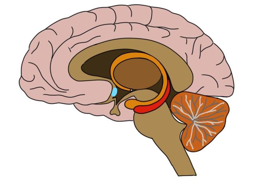
Functionally, the preoptic area is considered to be a region of the hypothalamus even though its embryological origins are as part of the telencephalon (rather than the diencephalon like the rest of the hypothalamus). It consists of the area of the hypothalamus that is situated at the very anterior (i.e. front) of the structure, and it extends back from the anterior of the hypothalamus to the posterior edge of the optic chiasm, the point where the optic nerves from the two eyes meet.
The preoptic area, like the rest of the hypothalamus, is a very functionally diverse region. This diversity is represented by the division of the preoptic area into a collection of distinct nuclei. The majority of the research into the preoptic area, however, has been done in rodents; it is not as well understood in humans. This has caused the anatomical differentiation of the preoptic area to not be completely consistent when applied to the human brain and has generated some disagreement, for example, as to which areas of the human preoptic area are homologous to those of the rat brain.
What is the preoptic area and what does it do?
As mentioned above, the preoptic area consists of several nuclei (each of which are often divided into subnuclei), and thus is a very functionally heterogeneous region. To attempt to simplify it, I will discuss the functions most commonly attributed to what are generally considered the major nuclei of the preoptic area (e.g. the median, periventricular, medial, and ventrolateral nuclei). The preoptic area is still not very well understood in humans, however, so the functions listed here will not be a comprehensive list of all that the preoptic area is involved in. Additionally, because most of the attempts to elucidate the function of the preoptic area has been done in rodents, it is still unclear if some of the functionality I will discuss can be said to describe the preoptic area in humans.
The median preoptic nucleus is found near the midline of the brain and at the very anterior end of the hypothalamus, where it borders the third ventricle. It merges and is neurally connected with a structure called the organum vasculosum, and it also receives input from another structure called the subfornical organ. The organum vasculosum and the subfornical organ are both part of a group of structures known as circumventricular organs. These structures lack a blood-brain barrierand thus can detect the levels of substances (e.g. sodium, hormones) in the blood, then pass this information on to the brain. The median preoptic nucleus is thought to receive such information from the organum vasculosum and subfornical organ and then seems to be involved in using that information to help regulate blood composition and volume, both through mechanisms of hormone release and through behavior like drinking.
The median preoptic nucleus also appears to play a role in the regulation of body temperature. Rodent studies suggest neurons in the median preoptic nucleus receive information regarding skin temperature and then send projections to neurons in the medulla that are involved in mechanisms that influence body temperature.
Just below the median preoptic nucleus is the preoptic periventricular nucleus. The preoptic periventricular nucleus is poorly defined anatomically (its boundaries vary depending on species) and poorly understood functionally. Some consider it to be equivalent to an area called the anteroventral periventricular nucleus, which is thought to be involved with sex-specific physiology and behavior, but according to other sources it is a separate location. There is limited information available on the preoptic periventricular nucleus as a stand-alone structure when it is not considered as part of the anteroventral periventricular nucleus or other nuclei nearby.
Lateral to the median preoptic nucleus is the medial preoptic nucleus. The medial preoptic nucleus is the largest collection of preoptic area neurons. It has been linked to a list of actions ranging from regulation of cardiovascular function to regulation of body temperature and fluid balance and water intake. This region, however, is best known for its association with reproductive and parental behavior.
The medial preoptic nucleus is often divided into subnuclei, and the central medial preoptic nucleus in the rat is sometimes called the sexually dimorphic nucleus, as it has been found to be larger in males than females. This observation has also been replicated in a number of other species. Although this has not been seen as consistently in humans, researchers have identified potential homologous regions in the human medial preoptic area that also have been observed to exhibit sexual dimorphism.
The sexually dimorphic nucleus in rodents has been linked to male sexual behavior and male partner preference. Lesions to different parts of this region have been found to eliminate male copulatory behavior and inhibit sexual desire. Additionally, damage to the sexually dimorphic nucleus in male ferrets was associated with their sexually-motivated seeking of same-sex males rather than females. In a study of sheep (which are unique in that ~8% of rams display a consistent preference for male sexual partners), it was found that the sexually dimorphic nucleus was twice as large in female-oriented rams than in male-oriented (i.e. homosexual) rams.
These findings, of course, have led to speculation that the sexually dimorphic nucleus in humans may also be linked to sexual preference. Although there has been some debate as to what the human homolog of the rat sexually dimorphic nucleus actually is, there have been studies that have found sexual dimorphism among preoptic regions in humans. Some studies have also observed differences in size in these regions between heterosexual and homosexual men. These findings, however, have not been seen consistently from study to study, and need to be confirmed before we can be confident in them.
Additionally, the central medial preoptic nucleus has been linked to parental behavior in rodents, sheep, and other mammals. For example, damage to the connections of the medial preoptic nucleus can disrupt parental behaviors like nest building and pup retrieval (i.e. collecting stray pups when they wander from the nest) in rodents. And activation of medial preoptic neurons mitigates male aggression toward pups as well as stimulates the grooming of pups.
Just above the optic chiasm is another nucleus that is known as the ventrolateral preoptic nucleus in rodents. It is generally considered to be homologous with a nucleus in the human brain called the intermediate nucleus, and this cell group actually overlaps with the nuclei discussed above that are thought to be sexually dimorphic in the human brain. This region is thought to play an important role in sleep regulation. Damage to the ventrolateral preoptic nucleus can cause sleep disruptions in a variety of mammalian species. One hypothesis is that ventrolateral preoptic neurons extend to brain regions involved in arousal; through the release of inhibitory neurotransmitters like GABA, the ventrolateral preoptic nucleus can inhibit the activity of these regions to promote sleep. The median preoptic area (discussed above) is also thought to potentially contribute to the induction of sleep.
The preoptic area consists of a collection of nuclei that are interconnected with numerous other hypothalamic regions and other regions of the brain. The anatomy and function of the preoptic area is still being worked out, and the functions presented here are just a short and very abridged list of everything the preoptic region is likely involved in. Additionally, due to the inconsistency in definitions of preoptic nuclei in the human hypothalamus, it is possible some of the definitions discussed above will change with further research into the preoptic area.
Reference (in addition to linked text above):
Saper, BS. (2012). Hypothalamus. In JK Mai and G Paxinos (Eds.) The Human Nervous System, 548-583 DOI: https://.org/10.1016/B978-0-12-374236-0.10016-1